
This manual guides users through the intricacies of anatomy and physiology, offering step-by-step procedures and access to over 6,000 clinical articles.
A. Lab Safety Protocols
Prior to commencing any laboratory work, a thorough understanding of safety protocols is paramount. This includes proper handling of specimens, appropriate disposal of biological waste, and adherence to all instructor guidelines. Always wear appropriate personal protective equipment (PPE), such as gloves and eye protection, to minimize exposure risks. Familiarize yourself with the location of safety equipment, including eyewash stations and first aid kits. Report any accidents or spills immediately to ensure a safe learning environment for everyone involved in the anatomy and physiology lab.
B. Use of Microscopy
Microscopy is a fundamental skill in anatomy and physiology, enabling visualization of cellular structures. Proper handling and maintenance of microscopes are crucial for optimal performance. Begin with the lowest power objective lens and gradually increase magnification, carefully focusing on the specimen. Understand the function of each component, including the condenser and diaphragm, to adjust illumination. Always clean the lenses after use with lens paper to prevent damage. Accurate observation and documentation are key to successful microscopic analysis.
C. Anatomical Terminology & Body Planes
A standardized anatomical vocabulary is essential for precise communication. Understand directional terms like superior, inferior, anterior, and posterior. Master the body planes – sagittal, frontal (coronal), and transverse – to accurately describe sections and dissections. Regional terms delineate specific body areas, aiding in localization of structures. Proper use of terminology ensures clarity in lab reports and discussions. Consistent application of these concepts builds a strong foundation for understanding anatomical relationships.

II. Histology: The Study of Tissues
Explore microscopic tissue structures, crucial for understanding organ function. This section details epithelial and connective tissue types, forming the basis of histological analysis.
A. Epithelial Tissue Types & Functions
Epithelial tissues cover body surfaces, lining organs and forming glands. This lab explores squamous, cuboidal, and columnar epithelium, examining their roles in protection, secretion, absorption, and filtration.
Simple epithelia facilitate easy passage, while stratified epithelia provide robust defense. Pseudostratified columnar epithelium, often ciliated, lines the respiratory tract. Transitional epithelium, found in the urinary system, allows for stretching.
Understanding these diverse types is fundamental to comprehending organ-level physiology and pathology, linking structure directly to function within the body.

B. Connective Tissue Types & Functions
Connective tissues support, connect, and separate different types of tissues and organs in the body. This lab focuses on connective tissue proper – loose (areolar, adipose, reticular) and dense (regular, irregular, elastic) – alongside specialized types like cartilage, bone, and blood.
Adipose tissue stores energy, cartilage provides flexible support, and bone offers rigid structure. Blood transports nutrients and waste.
Examining fiber types (collagen, elastic, reticular) reveals how structure dictates function, crucial for understanding tissue repair and overall body integrity.

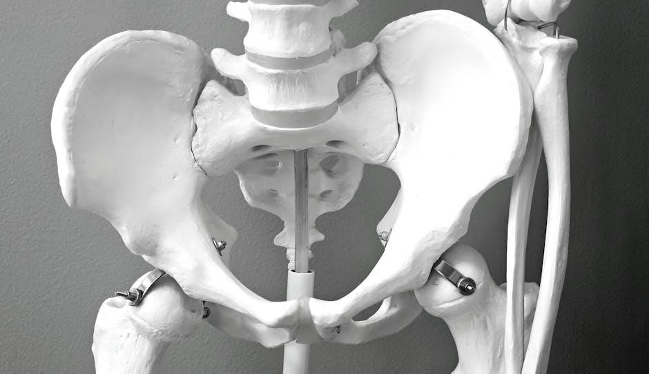
III. The Skeletal System
This section explores bone structure, classifications, and articulations, detailing joint types and their associated movements for comprehensive skeletal understanding.
A. Bone Structure & Classification
Understanding bone structure is fundamental; bones comprise compact and spongy tissue, with classifications based on shape – long, short, flat, irregular, and sesamoid. This lab manual provides detailed insights into the microscopic and macroscopic anatomy of bone, exploring features like the periosteum, endosteum, and various bone cells (osteoblasts, osteocytes, osteoclasts).
Furthermore, it elucidates how these structural components contribute to bone’s functions: support, protection, movement, mineral storage, and blood cell formation. Practical exercises will reinforce identification and classification skills, crucial for anatomical comprehension.
B. Articulations (Joints) – Types & Movements
Joints, or articulations, are critical for skeletal movement, categorized structurally as fibrous, cartilaginous, or synovial. This lab manual details each type, emphasizing their unique characteristics and range of motion. Synovial joints – ball-and-socket, hinge, pivot, saddle, plane, and condyloid – are explored with practical exercises. Students will learn to identify joint movements like flexion, extension, abduction, adduction, rotation, and circumduction.
Understanding these classifications and movements is essential for analyzing biomechanics and diagnosing musculoskeletal conditions.

IV. The Muscular System
This section of the manual explores skeletal, smooth, and cardiac muscle tissues, detailing contraction mechanisms and their physiological roles within the body;
A. Muscle Tissue Types (Skeletal, Smooth, Cardiac)
The lab manual meticulously details the three primary muscle tissue types: skeletal, smooth, and cardiac. Skeletal muscle, responsible for voluntary movements, exhibits a striated appearance under microscopy. Smooth muscle, found in the walls of internal organs, facilitates involuntary functions like digestion.
Cardiac muscle, exclusive to the heart, demonstrates both striated characteristics and involuntary control, ensuring rhythmic contractions. The manual provides comparative analyses of their structural differences, cellular compositions, and functional roles within the human body, aiding in comprehensive understanding.
B. Muscle Contraction Mechanisms
This section of the lab manual thoroughly explores the intricate mechanisms driving muscle contraction. It details the sliding filament theory, explaining how actin and myosin interact to generate force. The role of calcium ions, ATP, and nerve impulses in initiating and sustaining contractions is meticulously outlined.
Furthermore, the manual clarifies the steps involved in excitation-contraction coupling, emphasizing the neuromuscular junction’s crucial function. Diagrams and illustrations enhance comprehension of these complex physiological processes, providing a robust foundation for understanding muscular function.

V. The Nervous System
The lab manual details neuron structure and function, alongside comprehensive brain anatomy, including lobes, for a complete neurological understanding.
A. Neuron Structure & Function
This section of the anatomy & physiology lab manual meticulously explores the foundational units of the nervous system – neurons. It provides detailed illustrations and explanations of neuronal components, including the cell body (soma), dendrites, axon, and myelin sheath.
Students will learn about the crucial role of each structure in transmitting electrical and chemical signals. The manual further delves into the functional aspects of neurons, covering action potentials, synaptic transmission, and the influence of neurotransmitters.
Practical exercises and diagrams reinforce understanding of how these complex processes enable communication throughout the nervous system, forming the basis for all neurological functions.
B. Brain Anatomy & Lobes
The anatomy & physiology lab manual dedicates a comprehensive section to the intricate structure of the human brain. Detailed diagrams and 3D models illustrate the major brain regions, including the cerebrum, cerebellum, and brainstem, providing a clear visual understanding.
This module specifically focuses on the four lobes – frontal, parietal, temporal, and occipital – outlining their distinct functions and interconnectedness. Students will explore the roles of each lobe in higher-level cognitive processes, sensory perception, and motor control.
Interactive exercises and case studies enhance learning and application of this vital neurological knowledge.

VI. The Cardiovascular System
The lab manual details heart anatomy, blood flow, and vessel structure, utilizing clinical articles and step-by-step procedures for comprehensive study.
A. Heart Anatomy & Blood Flow
This section of the anatomy & physiology lab manual meticulously explores the heart’s complex structure, detailing chambers, valves, and major blood vessels.
Students will trace the pathway of blood flow through the heart, understanding systemic and pulmonary circulation with detailed diagrams and procedures.
Access to over 6,000 peer-reviewed clinical articles enhances understanding, while step-by-step guides facilitate dissection and identification of key anatomical features.
The manual emphasizes the functional relationship between structure and blood flow, preparing students for advanced cardiovascular concepts.
B. Blood Vessels – Structure & Function
This lab manual component delves into the intricate world of blood vessels – arteries, veins, and capillaries – examining their unique structural adaptations.
Students will analyze how these structures directly correlate with their specific functions in transporting blood throughout the body, utilizing detailed illustrations.
The manual provides access to clinical articles, enhancing comprehension of vascular diseases and their impact on circulatory efficiency.
Step-by-step procedures guide students through microscopic examination and identification of vessel layers.
VII. The Respiratory System
Explore respiratory structures and ventilation techniques with this manual, accessing clinical articles and learning units for a comprehensive understanding.
A. Respiratory Structures & Ventilation
This section of the anatomy & physiology lab manual meticulously details the complex respiratory system. It provides a thorough examination of key structures, including the nasal cavity, pharynx, larynx, trachea, bronchi, and alveoli, emphasizing their individual roles in the ventilation process.
Users will gain practical insights into the mechanics of breathing, exploring inspiration and expiration, alongside the muscles involved in these vital functions. Access to over 6,000 peer-reviewed clinical articles enhances understanding, while step-by-step procedures solidify learned concepts.
B. Gas Exchange Mechanisms
The anatomy & physiology lab manual dedicates a crucial section to understanding gas exchange, the cornerstone of respiratory function. This exploration delves into the intricate processes occurring within the alveoli and capillaries, focusing on oxygen and carbon dioxide diffusion.
Users will analyze factors influencing gas exchange efficiency, benefiting from access to over 6,000 clinical articles and detailed, step-by-step procedures. This section reinforces comprehension of partial pressure gradients and their role in maintaining respiratory homeostasis.

VIII. The Digestive System
The lab manual explores digestive organs and functions, alongside absorption processes, utilizing clinical articles and step-by-step guides for comprehensive understanding.
A. Digestive Organs & Functions
This section of the anatomy & physiology lab manual meticulously details each digestive organ, from the mouth to the anus, outlining their specific roles in the breakdown and absorption of nutrients.
It provides a comprehensive overview of the stomach’s churning action, the small intestine’s villi for nutrient uptake, and the large intestine’s water absorption capabilities.
Furthermore, the manual incorporates clinical relevance through access to peer-reviewed articles, enhancing understanding of digestive disorders and their physiological basis.
Step-by-step procedures aid in visualizing and comprehending the complex interplay of these organs, fostering a deeper appreciation for digestive system functionality.
B. Digestive Processes (Absorption, etc.)
The anatomy & physiology lab manual thoroughly explores digestive processes, including mechanical and chemical breakdown, enzymatic action, and the crucial stages of absorption.
It details how carbohydrates, proteins, and fats are processed, emphasizing the role of specific enzymes and the intestinal lining’s absorptive mechanisms.
Clinical articles within the manual provide context for absorption disorders and related pathologies, bridging theoretical knowledge with real-world applications.
Step-by-step guides facilitate understanding of these complex processes, enhancing comprehension of nutrient transport and utilization within the body.

IX. The Urinary System
This lab manual details kidney structure, urine formation, and regulatory functions, utilizing clinical articles and step-by-step guides for comprehensive understanding.
A. Kidney Structure & Function
The anatomy and physiology lab manual meticulously explores the kidney’s complex structure, from the outer cortex to the inner medulla, detailing the nephron’s crucial role in filtration.
It clarifies the processes of glomerular filtration, tubular reabsorption, and secretion, essential for maintaining fluid and electrolyte balance.
Furthermore, the manual provides access to peer-reviewed clinical articles, enhancing understanding of renal physiology and potential pathologies.
Step-by-step procedures aid in visualizing and comprehending the kidney’s vital functions within the urinary system.
B. Urine Formation
The anatomy and physiology lab manual comprehensively details urine formation, beginning with glomerular filtration – a process driven by blood pressure within the renal corpuscle.
It elucidates tubular reabsorption, where essential substances return to the bloodstream, and tubular secretion, eliminating waste products.
Access to over 6,000 clinical articles provides context for understanding how these processes are affected by various physiological conditions.
Step-by-step guides enhance comprehension of the intricate mechanisms involved in creating urine.